A dentist rotates a three-dimensional jaw on screen, tracing the relationship between a tooth root and the nerve canal.
AI is already entering different parts of this work. Sometimes it helps create the image itself; elsewhere, it picks out anatomical structures, places measurement markers or draws attention to a suspicious area. For the patient, that difference can mean less time in a scanner, a clearer plan for a procedure or one more check before going home. Let us follow that path, from teeth and bones to the beating heart.
A tooth in three dimensions, a fracture on an image
Structures overlap in a two-dimensional image. Dental CBCT, a form of computed tomography that produces a three-dimensional view, allows the jaw to be examined from different angles. The AI tool CEPHX3D separates out the upper and lower jaws, individual teeth with their crowns and roots, and the path of the inferior alveolar nerve. The dentist can rotate the model, adjust its transparency, measure distances and correct the boundaries marked by the software. An FDA document from March 2026 describes precisely this kind of assistance with analysis and planning.
The change is tangible: what the clinician once assembled mentally from a series of cross-sections becomes a spatial model they can manipulate. The relationship between root, bone and nerve canal can also be shown to the patient. A conversation about a procedure gains a shared image.
With an injury, the task is different. The X-ray is in front of the doctor, and software such as BoneView helps identify a possible fracture. In November 2025, Northern Ireland’s Department of Health described its use across four health trusts, through the existing system for viewing medical images. Under NICE guidance, four such tools can be used in the NHS while further evidence is gathered. In the consulting room, this provides another reason to look more closely at a particular area, alongside examining the patient and understanding how the injury happened.
Ten fewer minutes in the tunnel
With magnetic resonance imaging — MRI — help can begin before the radiologist receives the finished image. While the scanner collects signals to show the brain, spine or other tissues, the patient’s task is to stay still. A minute feels different to someone lying comfortably and someone in pain or struggling with an enclosed space.
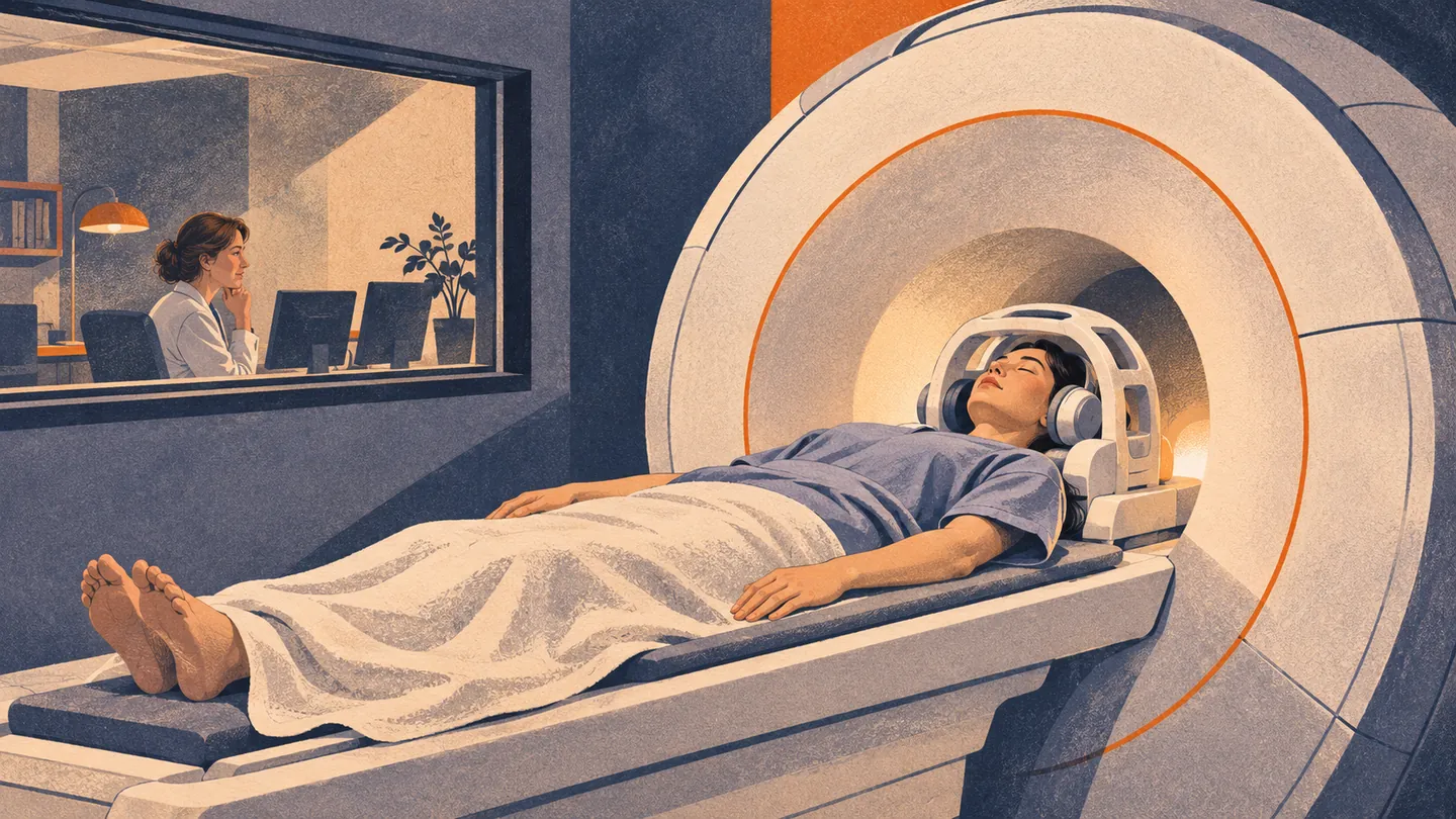
In July 2025, NHS Humber reported that introducing and upgrading AIR Recon DL software had shortened routine head MRI examinations in its practice from 30 to 20 minutes. The algorithm assists image reconstruction by reducing image noise, allowing quality to be maintained or improved with shorter data acquisition for certain scans. The team also describes people who can now get through an examination they previously found difficult. Ten fewer minutes of lying motionless is a concrete benefit for them.
There is a similar role in CT. Philips’ Precise Image uses deep learning in reconstruction to reduce noise and support usable images at a lower radiation dose, according to the examination and its protocol settings. Here, AI helps produce what the doctor will look at. Image quality, time and the burden on the patient become connected parts of the same decision.
While the examination is still under way
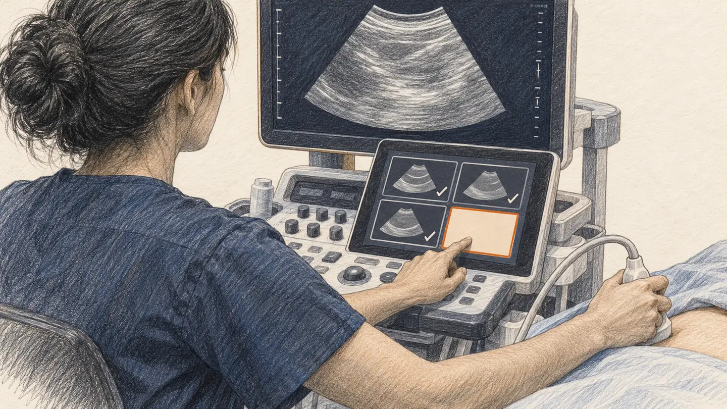
During an ultrasound examination, the image keeps changing. The probe moves, the angle shifts, and a detail that was hidden a moment ago comes into view. The doctor or sonographer finds the required views, records them and connects them with what they already know. For the person beside the screen, it is one examination; for the person performing it, a series of judgements to assemble into a whole.
With the heart, movement matters too: how the muscle contracts and how blood passes through the heart and blood vessels. Doppler provides information about flow, and AI can help clinicians work with those views. With Philips’ Auto Measure tool, a healthcare professional selects a standard 2D or Doppler measurement of an adult heart, and AI suggests where to place the measurement markers. The professional can move them, accept the result or reject it. The description of this function shows how much help can fit into a small, frequently repeated step of an examination.
For the fetal heart, the next question comes even earlier in that sequence: do we have all the required views while the probe is still in hand? This is where BrightHeart enters the story.
BrightHeart offers help at two related points. Its B-Right Views tracks whether the required views are complete, while B-Right Screen flags structural signs that may indicate severe congenital heart defects. Results can appear on a tablet beside the ultrasound machine while the examination is still in progress and further images can be taken.
The system shows which view has been recorded and which is still needed. The FDA document for the view-recognition module describes it as support for acquiring and interpreting ultrasound images, intended for trained healthcare professionals.
BrightHeart is already in use: Mount Sinai describes its introduction in 2025 at Carnegie Imaging for Women, which has three locations in Manhattan.
What the evaluations found
A retrospective analysis of 1,449 examinations at one centre was presented at SMFM 2026. After AI support was introduced, the share of examinations with all required cardiac views recorded rose from 59.5 to 91 per cent. Mean acquisition time did not change significantly. The analysis followed successive phases of the centre’s work, so it describes an improvement associated with introducing the system in that setting.
In a study published in Obstetrics & Gynecology, fourteen doctors reviewed 200 archived ultrasound examinations with and without AI assistance. Mean sensitivity — the ability to recognise suspicious signs when present — rose from 78.2 to 93.5 per cent, while interpretation time fell from 274 to 226 seconds. Half the selected examinations contained a suspicious finding, a much higher share than in routine screening; BrightHeart funded the study.
What else an existing scan may contain
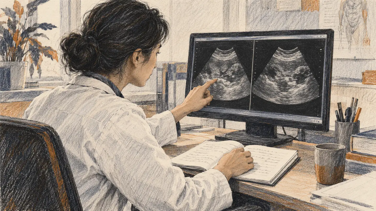
A CT examination has already been performed to answer one question. Imagine that the same scan offers a clue worth pursuing for another. That is the possibility being explored with Percival. Developed at the University of Pennsylvania, the model learns from scans and radiology reports. Researchers are investigating its ability to recognise patterns associated with disease and assess future risk. The paper was published in npj Digital Medicine in September 2026.
A scan performed for one reason might therefore offer a clue worth checking for another. According to Penn Medicine’s announcement of 16 October, the system is still in preclinical evaluation, and the team hopes to begin testing during 2027. How these signals could help in patient care remains to be investigated.
From image to sentence
After reading an X-ray, the findings need to be written up. Rology’s XGen prepares a draft chest X-ray report for a radiologist to check and complete. A conference analysis presented at RCR 2026 covered 2,824 examinations from Egypt, Kenya and Saudi Arabia. Here, assistance is being explored in report writing: what the radiologist keeps, what they correct and how the draft affects the final assessment.
When two readings meet
The doctor brings knowledge of anatomy and clinical context; the system brings patterns learned from many images. Their findings can complement each other. When they disagree, the flagged detail gives the doctor a reason to look again.
Why was the detail flagged? Is it visible in another view? Did the first impression guide the reading? Checking can confirm the initial finding or change it. Even a rejected suggestion may be useful if it helps the doctor explain the decision more clearly.
Across these examples, an additional look takes different forms. For one person, it means ten fewer minutes in an MRI scanner; for another, a spatial map of the jaw; for someone else, a closer check of a bone or a more complete heart examination. Its value grows when the clinician can compare their own observations with the system’s output and explain why they accepted, supplemented or rejected something.
For the patient, all of this must ultimately become an understandable conversation: what we saw, what it means and what comes next. Another look is worthwhile when it leads to a better next step.
Sources / Provenance
Additions checked on 3 November 2026: CEPHX3D based on FDA document K252538; BoneView based on Northern Ireland’s Department of Health, alongside NICE HTG739; AIR Recon DL based on NHS Humber’s experience reported in July 2025; Precise Image CT reconstruction and echocardiographic/Doppler Auto Measure based on Philips documentation. The figures for shorter MRI examinations are attributed to that institution. Earlier foundation, checked on 21 October 2026: BrightHeart through an image-reading study, a conference analysis of examination workflow, an FDA document and an account of clinical use; Percival through npj Digital Medicine and Penn Medicine; XGen through an RCR 2026 conference abstract. The introduction is an imagined walk through different examinations; the connections between examples and the encounter between clinical observation and AI analysis are editorial interpretation.